Diagnosis
Diagnosis is based on history and careful examination of the glottis across a variety of laryngeal tasks. Spasmodic dysphonia must be distinguished from other functional voice disorders such as voice tremor. An underlying neurologic disease must also be ruled out especially Wilson’s, Huntington’s and Parkinson’s disease which may cause secondary SD. Typical features of history include deterioration of vocal quality under stress or on telephone, and improvement with sedatives such as alcohol and benzodiazepines. Singing or laughing will sometimes result in greater fluency, probably due to the task-specific nature of dystonia. Diagnosis can be verified using electromyography, fiber optic laryngoscopy, videostroboscopy, aerodynamic testing, and vocal spectrographic analysis. Examination during connected speech is most likely to reveal the involuntary laryngeal motion that causes symptoms. That is why the larynx is best examined with a flexible nasopharyngoscope. Insertion of laryngeal mirror or rigid endoscope combined with the necessary traction of the tongue may mask the features. Diagnosis is based on speech symptoms and must be distinguished from functional voice disorder, and an underlying neurologic disease must be ruled out. The best evaluation involves a team approach and includes:
|
Electromyography
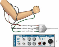
Electromyography is the recording of the electrical activity of muscle tissue, or its representation as a visual display or audible signal, using electrodes attached to the skin or inserted into the muscle.
Fiberoptic Laryngoscopy
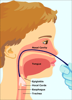
Flexible fiberoptic laryngoscopy utilizes a thin flexible endoscope that is inserted through the nose and passed into the throat under direct visualization. This provides a view of the structures of the throat, including the back of the nose (nasopharynx), the back of the mouth (oropharynx), the voice box (larynx), and the entrance to the swallowing passages (hypopharynx).
Videostroboscopy
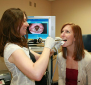
Videostroboscopy evaluates the function of the vocal cords, or larynx, in people with voice disorders. Stroboscopy uses a strobe light to illuminate the larynx. An examiner may then observe the movement and function of your larynx as you make particular sounds. This is done with either a flexible viewing tube (endoscope) passed through the nose or a rigid endoscope passed through the mouth. The scope contains an optical system and a tiny camera to record the exam on videotape for later review.
|
Differential Diagnosis
Clinical features can help distinguish between the different types of SD. Adductor type SD (ADSD) is found in about 85 percent of diagnosed cases in the United States. The most common symptom associated with adductor type SD is a choked, strained-strangled voice with abrupt breaks in phonation in the middle of vowels. Breaks are due to hyper adduction of the vocal folds resulting in a quick glottic closure interrupting airflow through the glottis and interrupting phonation. Patients may experience difficulties with continually voiced sentences particularly when glottal stops mark word boundaries like “we_eat,” or when two voiced sounds occur in sequence within the word such as “ye_ar” or “d_og”. Examples of sentences patients may have a difficulty with include “We eat eels every day”, “We mow our lawn all year” and “A dog dug a new bone”. Abductor type SD (ABSD) is less common, found in approximately 15 percent of patients with SD. Patients usually exhibit a breathy, effortful voice with abrupt breaks resulting in whispered elements of their speech characterized by excessive and prolonged abduction during voiceless consonants (/h/,/s/,/f/,/p/,/t/,/k/). Vocal fold abduction interferes with closure of the vowel sound that follows. To examine for symptoms of abductor SD the patient’s speech should be compared during voiced sentences such as “We mow our lawn all year,” which should contain few abnormalities, with sentences containing a high proportion of voiceless consonants such as “The puppy bit the tape” and “When he comes home we’ll feed him”. If severe enough, the patient may display complete aphonia. Mixed type SD is extremely rare. Patients display symptoms of both adductor and abductor type SD. Diagnosis of a mixed disorder is important for predicting response to treatment. Diagnosis is similar to the diagnosis of either type of SD, with patients having difficulties with both types of tasks. Mixed patients are difficult to treat as Botulinum toxin can produce unwanted side effects with no benefit. Thyroarytenoid injection produces breathiness that exacerbates the disorder, while injection to the posterior cricoarytenoid muscle may provide little benefit.
Sources:
Revelo, O. (2009). Spasmodic dysphonia: Evaluation and management. Grand Rounds Presentation, UTMB, Dept. of Otolaryngology. Retrieved from http://www.utmb.edu/otoref/grnds/dysphonia-090310/dysphonia-090310.pdf
ASHA. (n.d.). Spasmodic dysphonia. Retrieved from http://www.asha.org/public/speech/disorders/spasmodicdysphonia/
